Would you like to make this site your homepage? It's fast and easy...
Yes, Please make this my home page!
|
 |
William's (Liam's) May 28th Skydiving Accident
|
|

[New images are surrounded by a RED border]
(NOTE: Images and videos not related to Liam’s skydiving accident can be viewed at the bottom of this page… Enjoy!!)


(Click on the small images to see larger versions or play a video.)
(For videos it is best to right click on the video, select "Save Target As..." and then save the video file to your hard drive to be played.)
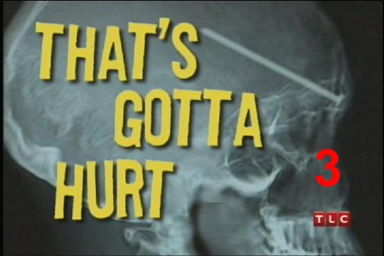
TLC Show "That's Gotta Hurt" Segments
The video clips below are captured from The Learning Channel (TLC) show which aired its first episode and first segment on February 14th 2007
covering Liam's May 28th 2005 skydiving accident. Since the full segment was too large to upload to the website as a single file, the show
has been broken up into 4 segments. Each file is a highly compressed Windows Media Video (WMV) formatted file with an estimated file size of
40 Meg per file. Download and viewing speeds may be reduced.
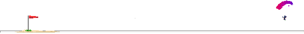
Videos of the actual landing (normal speed and supper slow-motion)
The two videos were captured from Liam’s helmet camera. The first video (left) is the real-time image of Liam going through the power lines.
On the second video (right) the video has been slowed down so that the viewer can see the cable contact at a frame-by-frame rate. The cable is
obviously coming at Liam in such a way as to possibly decapitate him. Liam was able to deflect the cable using his left arm. This action was
the only cognitive action that Liam took to protect himself.
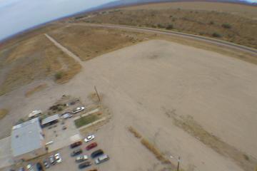
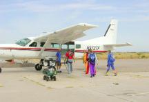
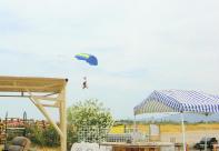
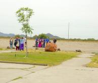
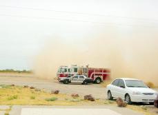
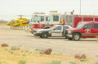
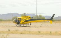
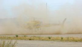
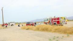
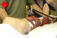
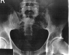
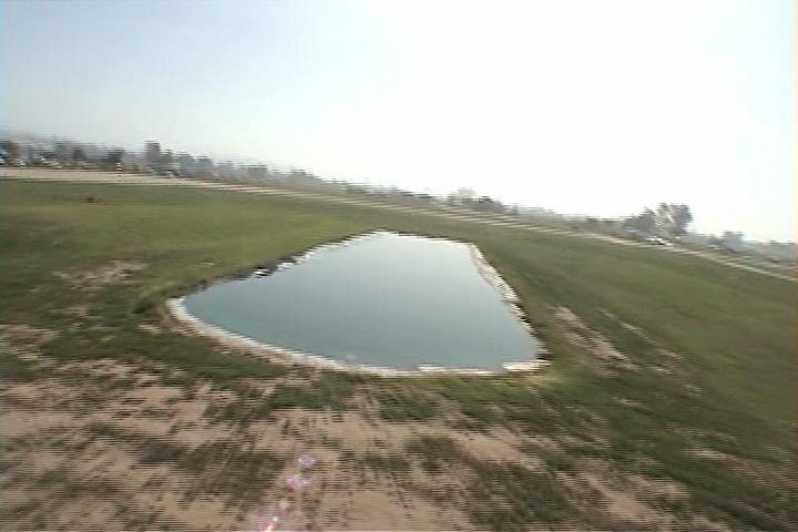
At the scene of the accident
These images show examples of skydiving routines (e.g. Loading the plane & Tandem landing). These images also show the various emergency
response crews that were called to address Liam’s accident. Liam remained on the ground (fully conscience) for an estimated 45 minutes
before being air lifted to St. Joe's Mecial Center in Phoenix. Liam was told that any other method of transportation would result in his death.
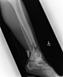
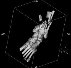
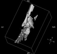
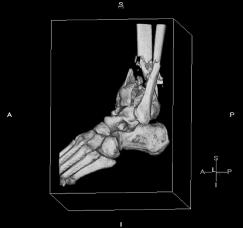
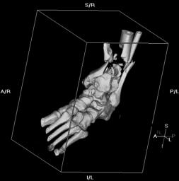
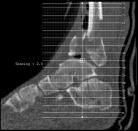
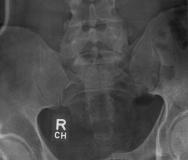
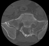
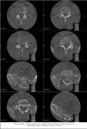
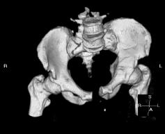
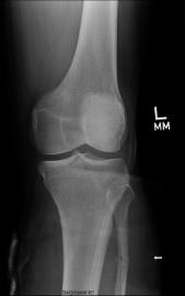
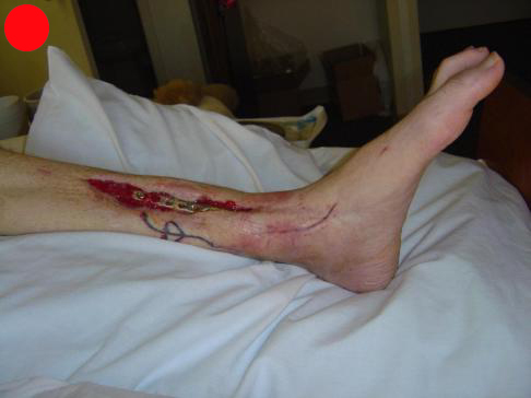
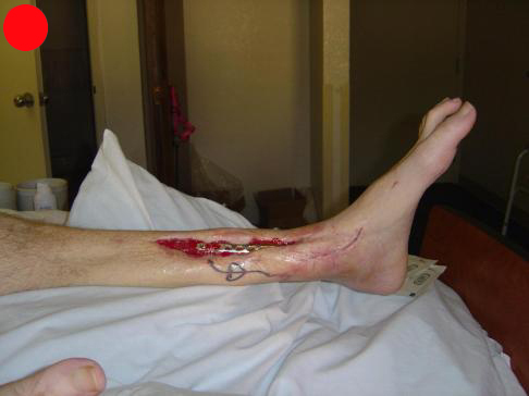
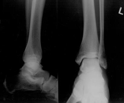
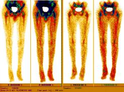
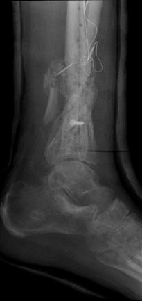
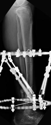
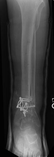
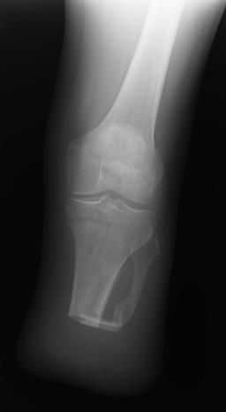
X-Rays and scans of the damaged right & left legs plus the pelvis
These x-rays were taken just after the accident. Some of the bones on the right leg have already been pushed back into the leg. The CT
scans show various views of the ankles and pelvis prior to any corrective surgery. Fibula bracing is clear in some of the images as well as
bracing that was used to secure the "open book" pelvis. In some of the last images it is apparent that the upper part of Liam’s left fibula is
broken. Liam was told that the pain he felt was most likely a muscle cramp. Nothing was done to repair this break and you will see in further
x-rays that the bone shifted and later fused in such a way as to add to the reduction of Liam’s leg length. The last photo shows the stint
used to block any potential blood clots from moving up.
[WARNING: Do not click on images marked with a red dot if you have a weak stomach]
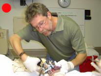
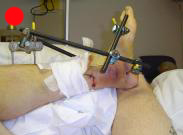
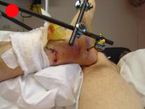
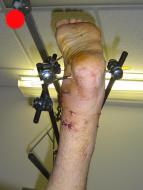
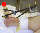
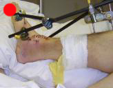
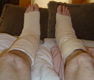
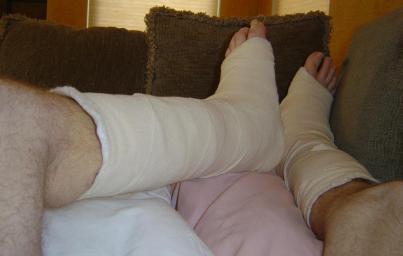
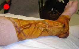
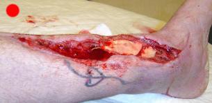
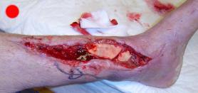
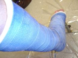
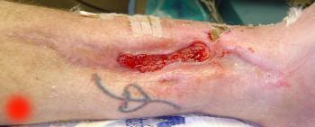
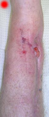
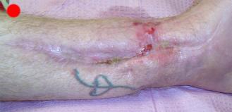
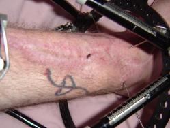
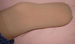
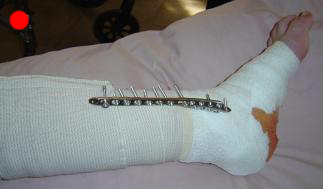
Hospital images of the damaged legs
These images were taken just after the first set of surgeries. The marks on Liam's face are from the riser strike. The external braces are
being used to allow the fibula of both legs to heal. Since there is no internal bracing provided to the tibias the bones are basically just
floating around in the leg (see x-rays in the "Pre-corrective surgery X-Rays of the damaged right & left legs / ankles" section) with the
hopes that they will not fuse in the wrong way. Liam was told at least 3 times that Dr. Keller would prefer to amputate the left leg.
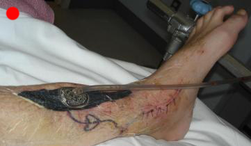
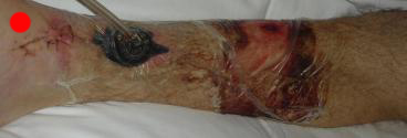
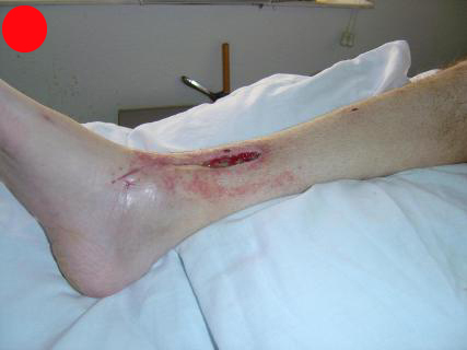
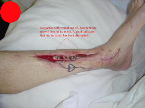
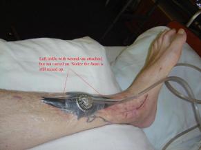
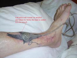
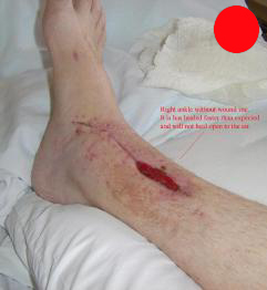
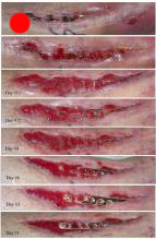
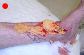
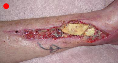
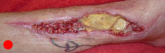
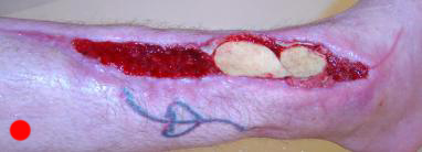
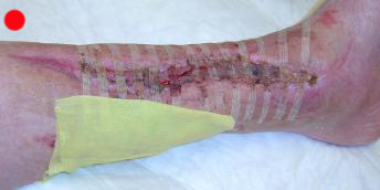
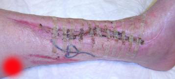
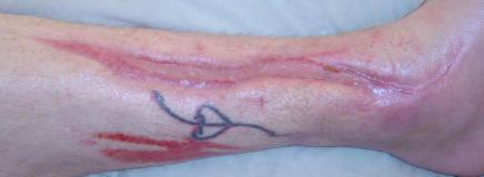
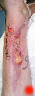
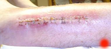
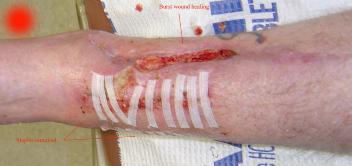
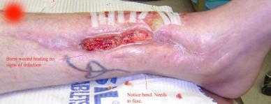
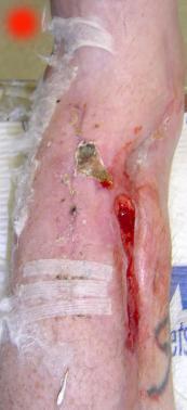
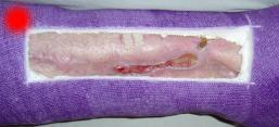
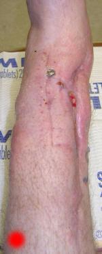
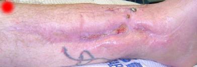
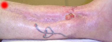
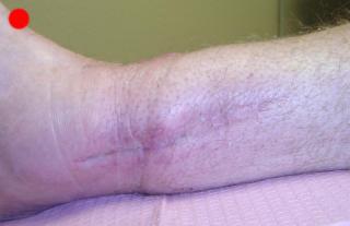
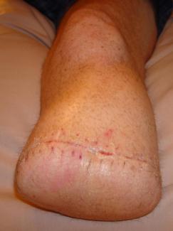
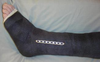
Hospital images of the damaged legs following corrective surgery (plus cast shots)
These images represent the various stages of healing that Liam’s legs were experiencing. In these images you can see the exposed plates
that are now in place over Liam’s tibias. These plates were not added during the procedures preformed by Dr. Greg Keller. That fact and many
more are the reasons that another doctor was selected to complete the surgeries on Liam’s legs. Notice the wound vacs used to promote healing
over the open wounds. The vacs were very good at doing their job. In a few images you can see the rate at which Liam’s wounds were healing.
The doctors were amazed at Liam’s healing rate. Some of the last photos show the left ankle post infective tissue / tibia plate removal. The
latest surgery was performed to debris Liam's tissue back to a level where tissue could grow or be grown over the wound. Tissue was removed
from another part of Liam's leg and placed over the wound. Dr. Gottlieb then infused bone material to promote bone growth and to close up
the open areas that existed in Liam's tibia. One of the photos is of a Pomegranate, since it was felt that Liam's leg looked like one. ;-)
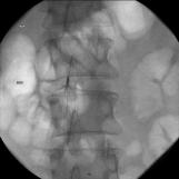
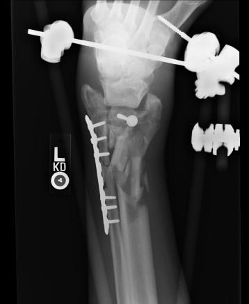
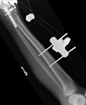
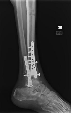
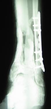
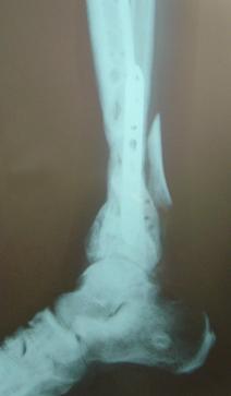
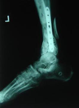
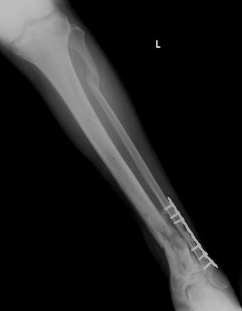
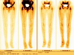
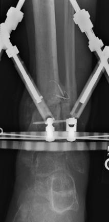
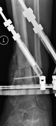
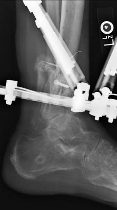
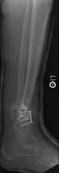
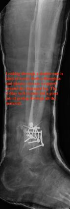
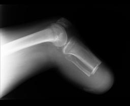
Pre-corrective surgery X-Rays of the damaged right & left legs / ankles
These x-rays were taken at Dr Armendariz’s office, just prior to performing any corrective surgery. As can be seen in these images,
there was no tibia bracing provided by Dr. Keller. Liam was released from Dr Keller’s care with instructions that full weight bearing could
be accomplished within 2 months of Keller’s last surgical procedure. The best example to examine is the second image (from the left) of the
top how. Notice how the bones that should be aligned with the tibia are in fact on the other side of the leg. The third image shows how badly
Liam’s left foot was twisted as a result of the pool placement of the external fixation. What is not obvious is that the screw at the bottom
of the plate on the right fibula missed being screwed into the plate.
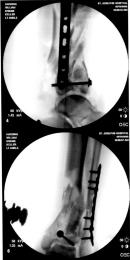
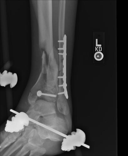
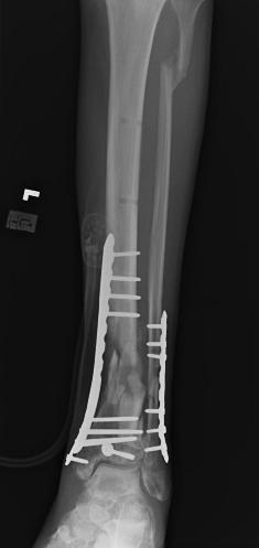
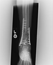
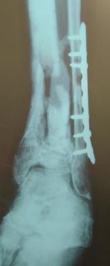
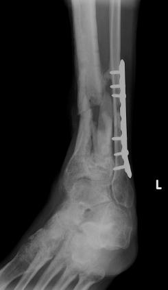
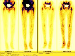
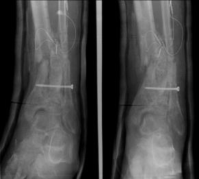
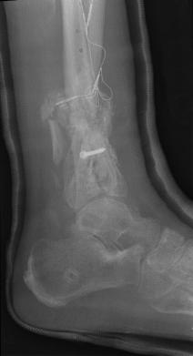
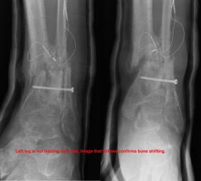
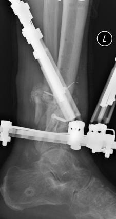
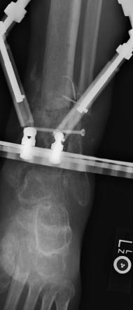
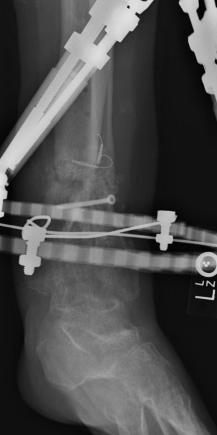
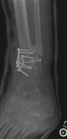
Post-corrective surgery X-Rays of the damaged right & left legs / ankles
The first two x-rays show Liam’s left ankle and pelvis prior to the accident. For comparison purposes it is interesting to see the
differences. The next x-rays were taken after the corrective surgery performed by Dr. Armendariz. Noticed that Dr. Armendariz has used tibia
bracing and also aligned the bone fragments of the left leg so that they could heal in the proper orientation. The external fixation has been
removed and Liam’s left foot has been returned to a more natural alignment.
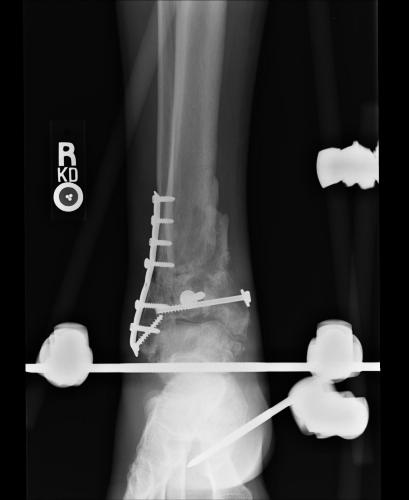
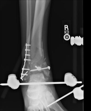
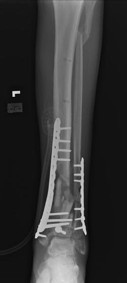
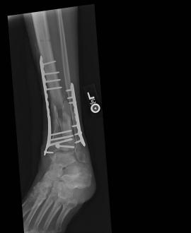
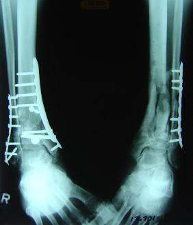
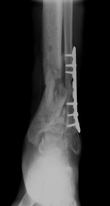
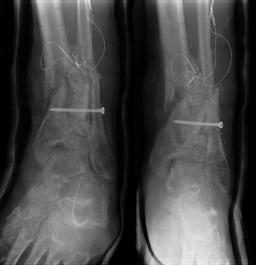
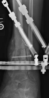
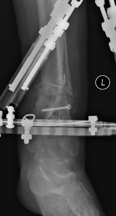
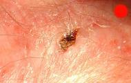
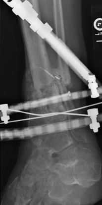
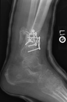
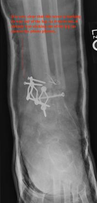
Post-infection X-Rays of the damaged right & left legs / ankles
After the last surgery Liam developed Staphylococcal (staph) infection as well as Osteomyelitis on the bone. Liam went to the ER of
Tempe St. Luke with a temperature of over 104 degrees and severe pain of the left leg. Liam was released from the ER with a diagnosis of
Gastroenteritis and instructions to follow up with a visit to Dr. Armendariz. Four days later and with a continued very high temp, Liam was
seen by Dr. Armendariz. Dr. Armendariz immediately performed 3 surgeries to clean out the infected areas of both the left and right leg and
to place wound vacs on the damaged areas of the legs.
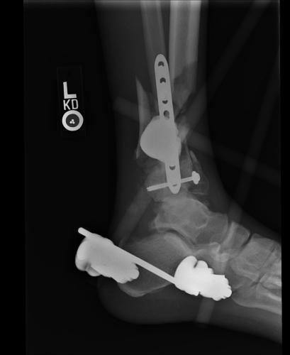
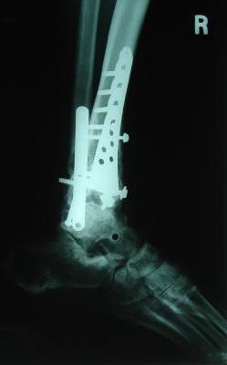
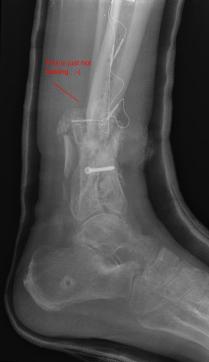
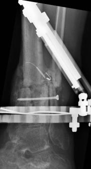
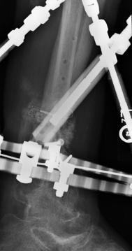
An interesting point to note, as it applies to the second and third x-rays (from the left), is the placement of the last screw at the bottom
of the right legs fibula plate. Notice how Dr. Keller completely missed the plate. Not only is the screw useless, but it has started protruding
in such a way as to cause severe pain and require padding to keep from bursting through the skin. A later surgery is planed for the screws
removal.
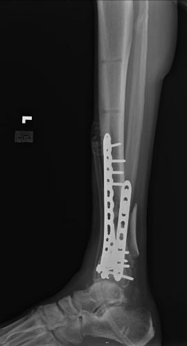
Pre-eighth surgery X-Rays of the left ankle
These x-rays were taken just prior to surgery for removal the left legs tibia plate and to perform a debrisment of the infected leg. Liam’s
left leg has not healed as well as the right leg and again Liam has developed Staphylococcal (staph) infection and Osteomyelitis on the bone
No x-rays were taken after the surgery and the only post operative images that can be viewed are in the "Hospital images of the damaged
legs following corrective surgery" section above (bottom five images).
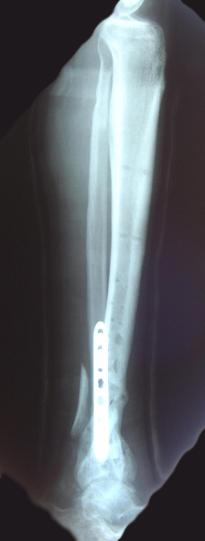
Pre-skin graft & bone infuse surgery X-Rays of the left ankle
These x-rays were taken just prior to surgery where grafts of skin (from Liam's own leg) were used to cover the open wound on Liam's left
ankle. Dr. Gottlieb also took the initiative to infuse material into the tibia gaps (shown in the front ankle view). The infused bone material
should fill in the existing bone gaps and fuse the various bone fragments, resulting in a much stronger support structure. It is hoped that the
3 cm fragment (shown in the side view) will fuse with the other tibia bones as time progresses. No immediate plan exist to modify the remaining
bone fragments / configuration.
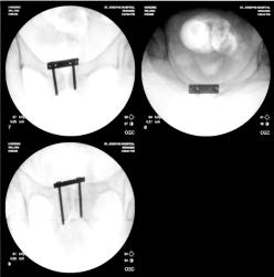
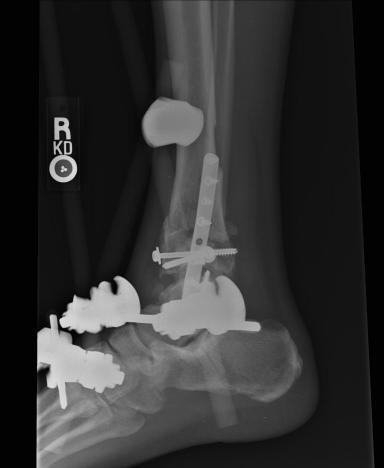
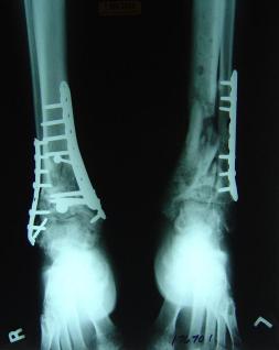
Post-skin graft & bone infuse surgery X-Rays of the left & right ankle
These x-rays were taken four weeks after surgery where grafts of skin were used to cover the open wound on Liam's left ankle. Dr. Gottlieb
infused bone material into the tibia gap of the left ankle. The 3 cm fragment in the left ankle does not appear to have fused or to be
obstructing normal movement so no immediate plans exist to modify the remaining bone fragments. Following these x-rays it is the plan to
remove the right ankle screw that was misplaced by Dr. Keller.
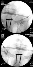
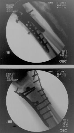
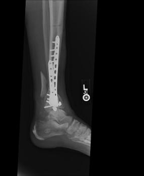
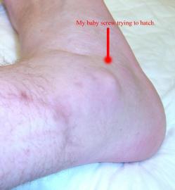
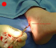
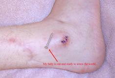
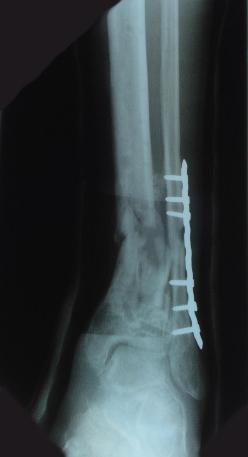
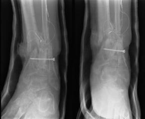
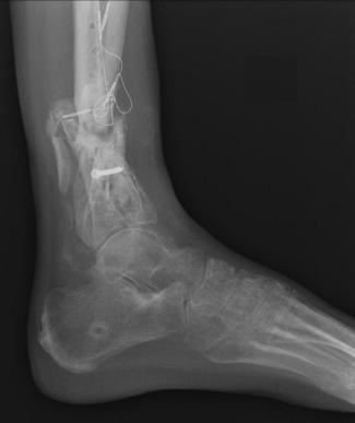
Screw Removed from Right Ankle
As can be noted in many of the right ankle x-rays, there was a screw that Dr. Keller placed in Liam’s lower right fibula which was
supposed to be through the plate in the fibula, but instead Dr. Keller missed the plate and simply screwed it into the bone. In the
position that the screw was place it caused considerable pain for Liam and obstructed Liam’s normal foot motion. The images below
show Dr. Gottlieb correcting Dr. Keller’s mistake.
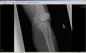
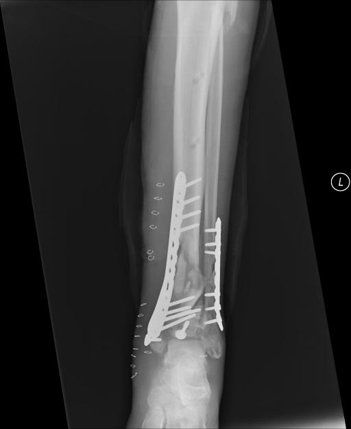
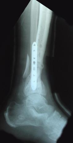
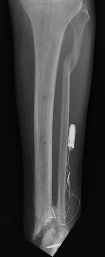
X-Rays Following Hyperextension of Left Ankle (January 31st 2006)
These x-rays were taken following a fluke event where Liam hyperextended his left ankle while playing an XBox 360 video game. As can be
seen in the images there are two (2) breaks that were produced from the hyperextension. One of the tibia bones has a hairline break and
the bone piece that spontaneously fused the tibia to the fibula has snapped. It took almost 2 weeks to determine that these breaks had
occurred. Due to Liam’s unique pain threshold and the lack of visible indicators, a break was not considered. Just another senseless
accident that resulted from the use of a violent video game. ;-) Yeah, right...
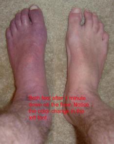
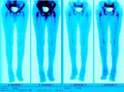
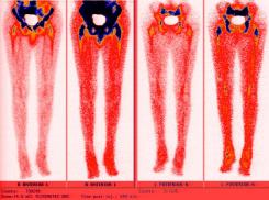
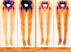
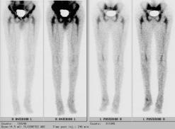
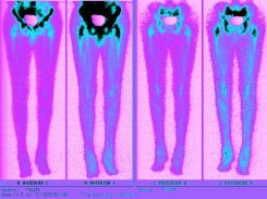
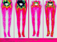
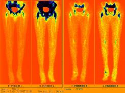
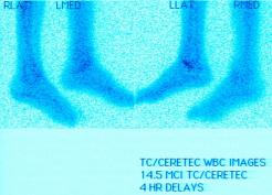
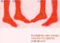
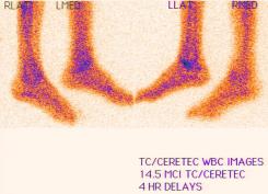
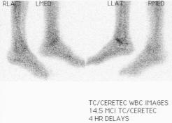
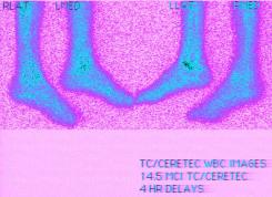
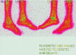
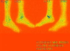
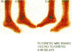
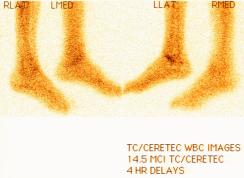
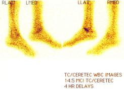
Nuclear Scan Images of Lower Torso & Lower Legs
These images were captured in order to determine why Liam’s bones broke so easily playing a video game and to also determine if there
was any Osteomyelitis. This is the diagnosis report from the scan. "There is mild increased activity noted in the lateral side of the left
ankle. However the patient does have a cast on the left leg. This uptake can be secondary to either inflammatory or infectious change within
either the skin or adjacent bone." Various contrast were used to enhance image details. The last images reminds one of the Shroud of Turin.
Hummm, makes one wonder... ;-)
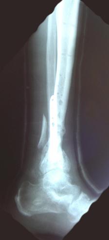
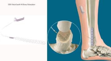
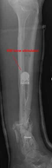
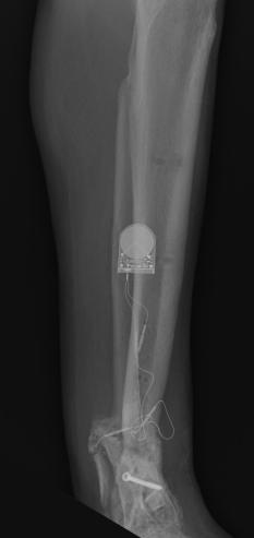
Preoperative X-Rays of the left ankle
These x-rays were taken just prior to surgery where Dr. Saunders planed to build up the left ankle with material, to repair any alignment issues
and to implant an electrical device that promotes bone growth and healing (like the EBI OsteoGen-M device shown in the last photo).
The ghost image around the leg is from the cast that was added following Liam's X-Box break. In some of the images, it can be seen
that Liam has removed the small window cover that was cut to allow access for the use of an Exogen ultrasonic bone-healing device.
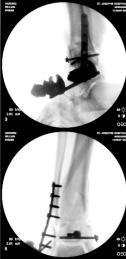
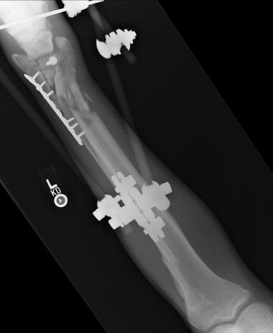
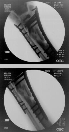
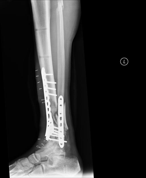
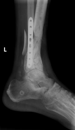
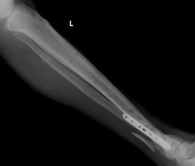
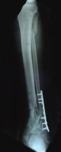
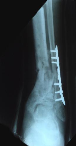
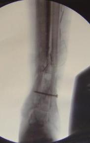
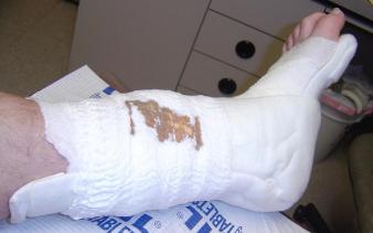
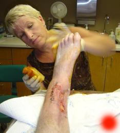
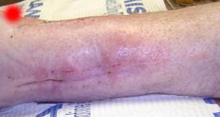
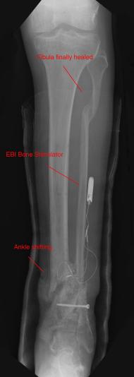
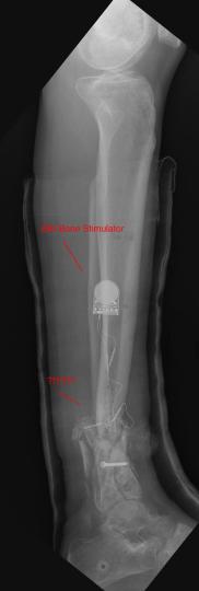
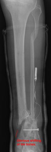
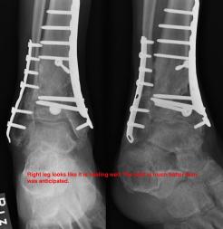
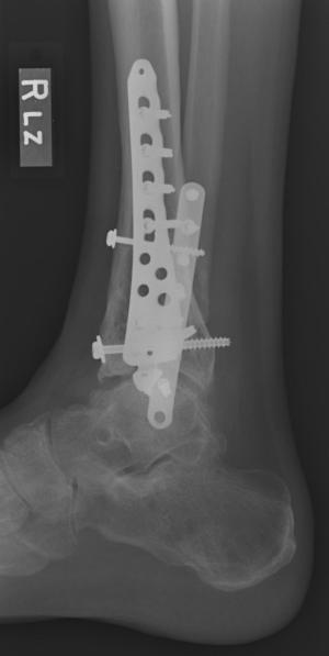
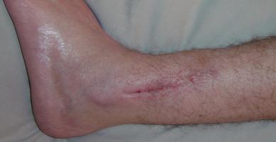
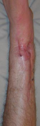
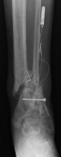
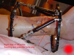
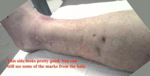
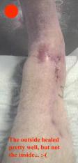
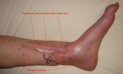
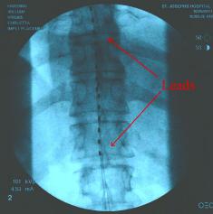
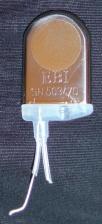
Postoperative (12th) images of left ankle
The images displayed here were created at various appointments following surgery performed by Dr. Daniel Saunders. In the X-Ray you can see
where Dr. Saunders has removed the fibula plate, joined the tibia and fibula with a screw, built-up the area of the tibia where previously
there was considerable absence of bone and lastly added the EBI OsteoGen-M Bone Stimulator (dual lead). The photos show the left side site
where the fibula plate was removed, the top site where donor bone, bone filler material and the stimulator were added and the right side
where the tissue has burst because of swelling. Liam was placed on an IV drip of Vancomycin following surgery to combat the potential onset
of Osteomyelitis. The image of the cast includes the fibula plate that was removed during surgery. The next images were taken later and show
that the staples and stitches were removed. In one of the images, you can see Liam's wife Madeline helping with leg cleanup and in one image
can be seen the EBI representative after being put to work (holding up the left leg while the latest cast cures) by Dr. Saunders. Later shots show
a healing tissue burst site with some overgrowth and Dr. Saunders cauterizing the overgrowth. Bones shifting too much (shown in X-Rays). The
last images are of the left ankle prior to the 13th surgery.
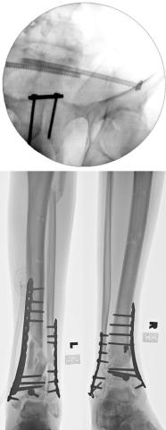
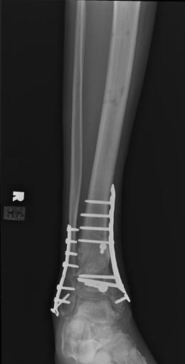
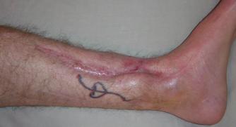
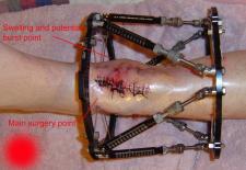
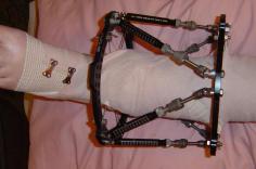
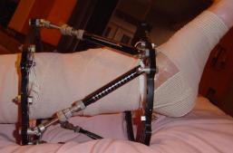
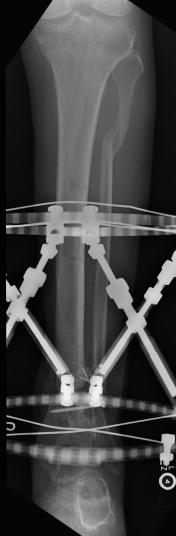
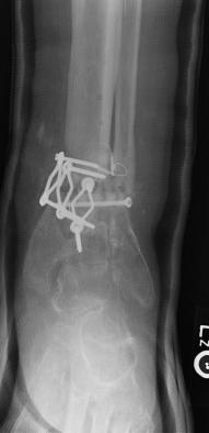
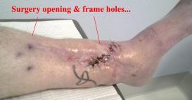
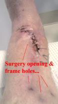
Postoperative (13th) images of left ankle
The image(s) displayed here were created following Liam's 13th surgery which was performed by Dr. Saunders. After a few hiccups getting
the surgery scheduled, Liam spent most of the September 13th afternoon in his 13th surgery. ;-) The surgery lasted about 5 hours and according
to the doctor, it was a good surgery. The 1st photo shows the new frame bracing that holds Liam's ankle together. The next series of photos show
various angles of the ankle and the rods that go from the frame into and through the left ankle. There was some concern that the swelling would
burst and allow for infection. Looks like we are now past that concern. Liam is still on antibiotics to ward of potential infection. The flesh
images show how the skin reacts to having the leg adjusted by the brace. The doctor has had to cut the skin to allow the rods to move freely.
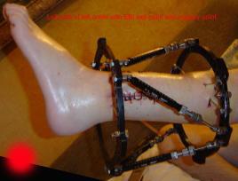
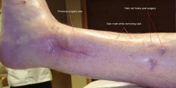
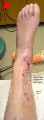
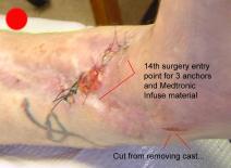
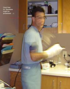
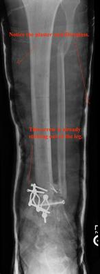
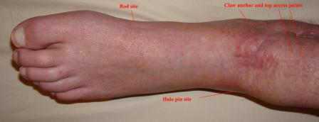
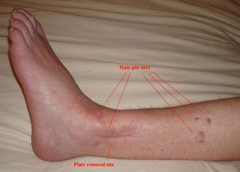
Postoperative (14th) images of left ankle
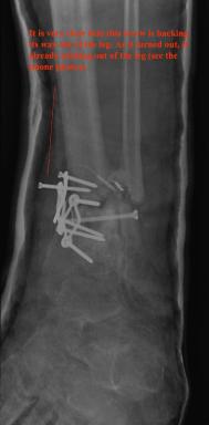
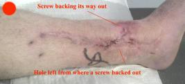
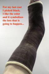
The image(s) displayed here were created following Liam's 14th surgery which was performed by Dr. Saunders. It was thought that the bones
in the ankle had fused, but during surgery it was discovered that the ankle was a total non-union. The halo frame was removed and 3 anchors were
placed on the tibia. Also added to the ankle was all available Medtronic Infuse material (rhBMP). One image may provide proof that Dr. Saunders
is possessed. ;-) The first set of x-ray shots were taken 3 weeks after the 14th surgery. The 3 new claw anchors are visible as seen through
the cast. Excess bone debris was also removed. Some of the photos had to be taking with the cell phone since the main camera failed. The last
x-rays show that some of the screws in the claw anchors are backing their way out of the leg. The next phone photos show that a screw from a
claw anchor was actually sticking out of the leg and could easily be removed (by hand) by the doctor. The very last photo is of the leg wrapped
in a black cast. Black was used to signify the loss that was soon going to occur. :-(
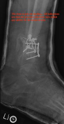
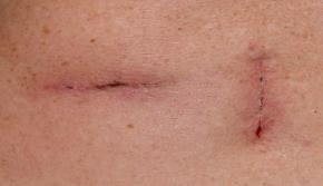
Preoperative (15th) images of left ankle
The following photos were taken 5 days before the amputation of Liam's left leg (below the knee). Liam has surrounded himself with a great team
which has allowed him to focus on work and not on the stress of the upcoming surgery. Of course leading that team is Madeline (Liam's wife). Madeline’s
dedication and support has been unwavering and the cornerstone of Liam’s strength. Note: The images shown were taken with Liam’s new 12.1 meg
digital camera following the CCD failure of Liam’s 5 meg camera.
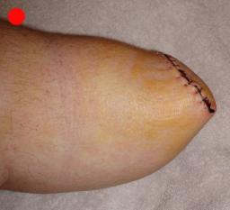
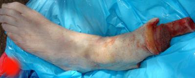
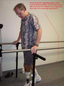
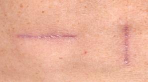
Postoperative (15th) images of left ankle
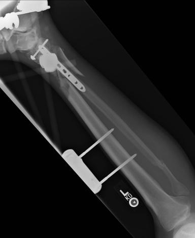
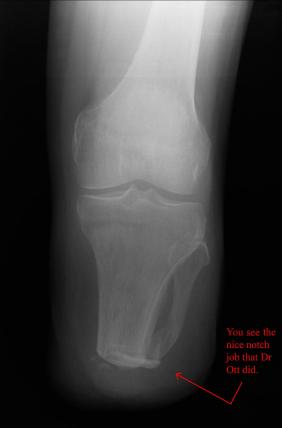
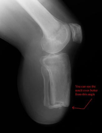
On May 30th 2007 exactly 2 years and 2 days from the day that Liam had his skydiving accident, doctor Ott removed the lower part of Liam’s left
leg using what is described as an Ertl procedure. The Ertl procedure results in a residual limb that is stronger and has a greater ability to load bear.
It is expected that Liam’s fatigue will be greatly reduced as a result of the Ertl procedure. The X-Rays show the best details associated with the Ertl
procedure. It can clearly be seen that a piece of bone (from the fibula) has been used to bridge the gap between tibia and fibula. What can not be seen
is the amount of tissue and muscle that was used (found normally in the lower part of the limb) to form a sort of pocket surrounding the remaining bone.
Through the great efforts of doctor Ott and Owen from the Artificial Limb Specialist Group, Liam has been given good responses regarding his rate of
healing and his prognosis for a great recovering. At this stage Liam is using sleeves that help reduce swelling and shape the stump so that it best fits the
prosthetic limb that will be used by Liam to walk.
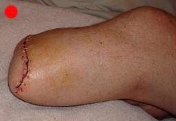
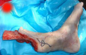
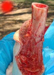
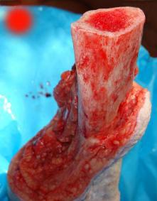
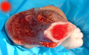
Amputated Leg Photos: Liam was able to reacquire his amputated leg from the Pathology lab. The lab diagnosis was “Infected nonunion left tibia”.
Basically after Liam’s leg became infected there was nothing that was going to allow Liam’s bones to knit. Liam transported the amputated leg to a
mortuary to have it cremated, but before delivering the leg Liam shot the last 7 images. Liam’s plan is divide the ashes so that some can be scattered
in a skydive over Buckeye and the remainder can be spread over the ocean.
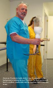
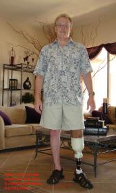
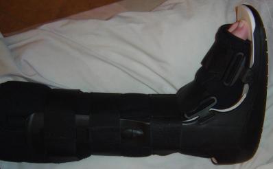
Liam's New Leg (Prosthesis)
After more than 2 years Liam is finally up and using a leg replacement for his amputated left leg. The foot is an Ossur FlexFoot LP Vari-Flex
and the pylon is a MedVentions Durashock. The socket is a clear polymer used to determine fit and until the final carbon fiber socket is prepared.
In these images Liam has had the prosthesis for about a week and still uses a single Canadian crutch for stability. Liam’s prosthetist “Owen” is also
shown in one of the photos.
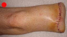
8 Months(+) After Amputation (Prosthesis)
Putting a little time between the amputation and Liam's acclimation to the prosthetic, these images are of the most recent x-rays of Liam's
left leg, which looks great and is able to support full body weight. Liam's is almost off all of the drugs and able control the residual pain. As can
be seen in the x-rays, Dr Ott did a great job with providing Liam with a load barring bone structure. The images of Liam's 2 prosthetic legs provide
an idea of what the legs are used for and their capabilities.
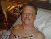
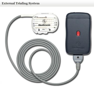
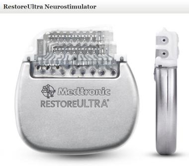
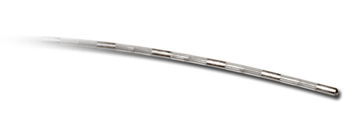
Surgery 16 (Pain Control)
After a number of years dealing with the pain resulting from the left leg below the knee amputation and damage that was residual in Liam’s
left buttocks (where his pelvis decelerated into the soft tissue), Liam decided that it was time to take a larger step in reducing the pain. Liam had
tried drugs as prescribed by his doctors, tried various vitamin injections into the damaged nerves (to repair those nerves), and Liam even tried
injections directly into the nerves along his spinal column, but nothing appeared to work for more than a few moments. So Liam had a Medtronic
RestoreUltra® implanted... Before the final implant was made Liam went through a device trial, to determine the feasibility of the final implant. The
RestoreUltra® is a multi-programmable, rechargeable neurostimulator that is part of a spinal cord stimulation system. The components that make
up Liam’s new toy, those used in the device trial, and some of the post-surgery photos can be seen in the following images.
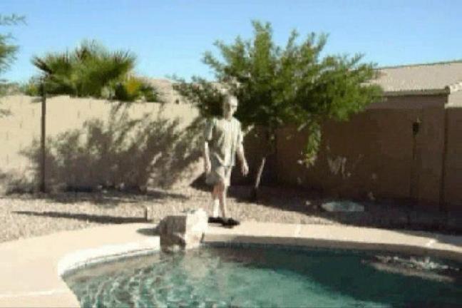
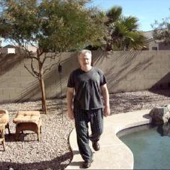
Walking Videos
The images show Liam’s progress towards returning to a normal life. The first walking video was taken days before the surgery that was
performed to remove the left legs tibia plate. The second walking video was taken 1 year and 3 months later and after considerably more surgery.
Considering that the doctors wanted to amputate Liam’s left leg and that it was never expected that Liam would walk again, these feats speak for
them selves.
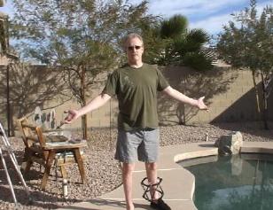
Post Amputation Videos: Eight months of Liam's leg was amputated it seems clear that he has returned to walking normally. Additional videos shot at the
physical therapy center give support to the fact that it has only taken Liam a short while to adjust to the prosthetic and is able to perform most ambulatory
actions with little difficulty. Liam continues to receive physical therapy and reports improved functionality in both legs.
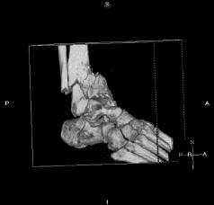
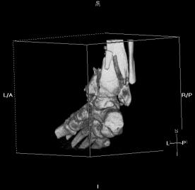
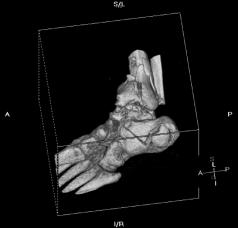
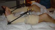
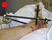
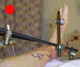
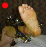
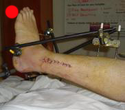
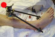
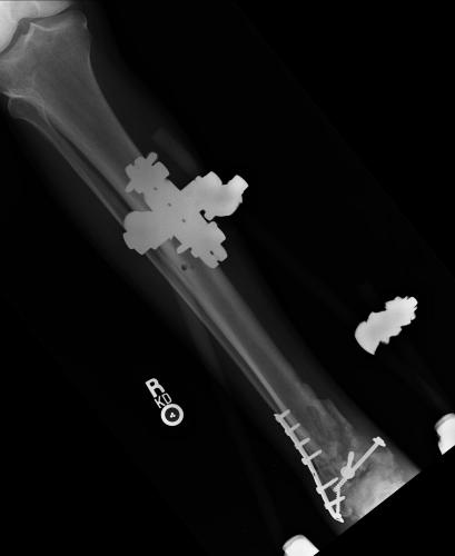
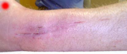
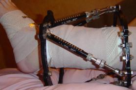
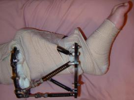
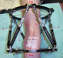
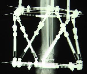
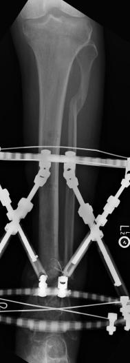
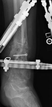
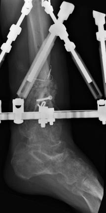
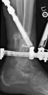
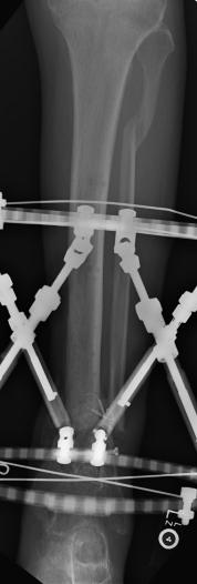
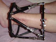
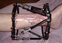
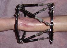
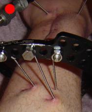
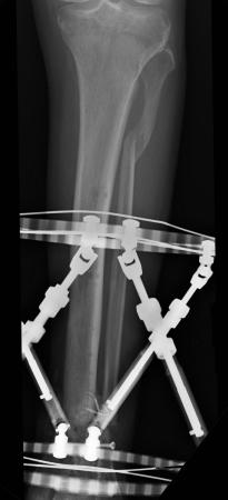
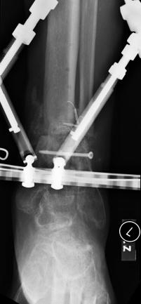
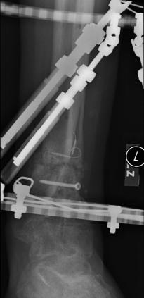
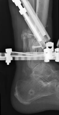
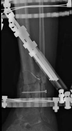
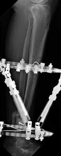
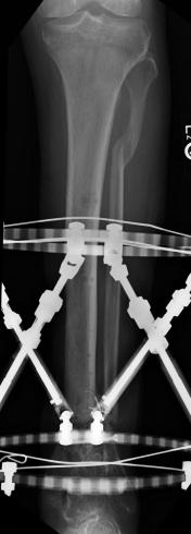
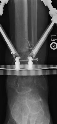
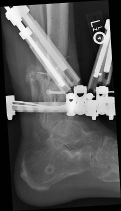
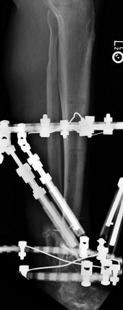
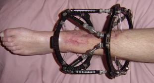
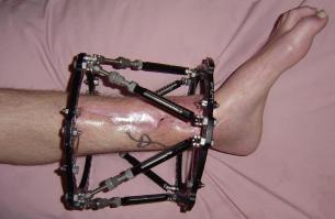
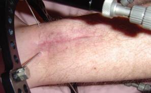
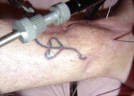
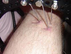
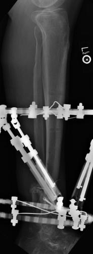
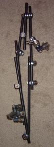
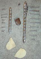
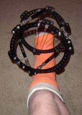
Fixation Components
The following images are of various fixation components. What is shown are some the components that were internal
to the left leg, some that were both external and also through the ankle and lastly some that were strictly external.
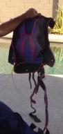
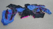
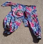
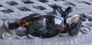
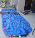
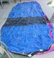
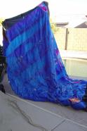
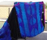
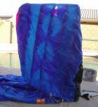
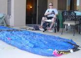
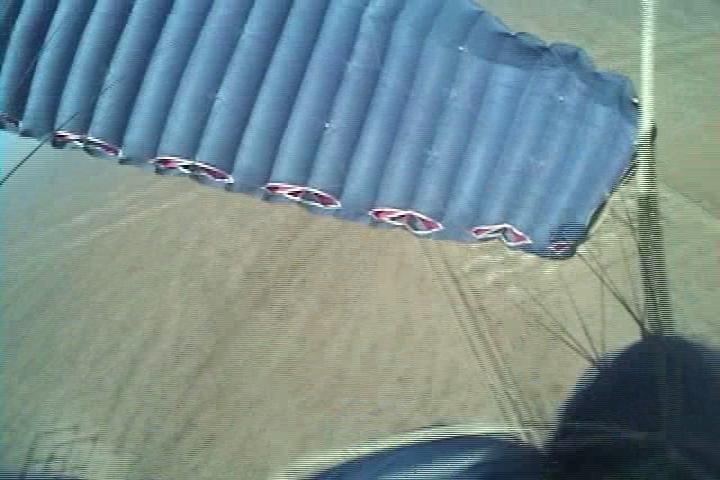
Photos of the damaged parachute and associated components
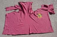
The photos below show the damage that Liam’s skydiving gear sustained from the accident. Liam’s clothing had to be cut off and thanks to
Liam’s skydiving friends, his container and helmet were protected from damage. Notice how the right side of the canopy (which was only a month
old) has had the lines cut. It is clear from the damage shown in the photos and the video footage (shown below) that as Liam’s body was swinging
up (after contacting the power lines), that his lines were being cut until such time as Liam’s body reach a angle where the canopy was released
from the power lines. The goggles show the marks that the power lines made as they rolled up Liam’s face.
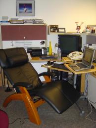
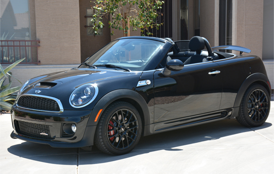
Post-accident changes
These photos reflect changes that have been made as a result of the accident. Because the damage to Liam's left leg and the fact that the Porsche
(Boxster S) had a clutch, it remained unused in the garage for most of the year. Liam got rid of the Porsche and ordered a (BMW) Mini (Cooper S
Convertible) and now enjoys driving a legal go-cart (as can be seen at the following YouTube video). Additionally, Madeline’s new BMW show’s
that the Harding’s are a 2 Bimmer family. Shown here also are changes that Liam's work made to his office (e.g. desk chair, removed island, etc).
YouTube "WCBH Highway 2 Hell"
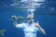
Post-accident life continues
The images that follow show that Liam has very effectively continued a normal life (ok... not really a normal life, since Liam is not normal). Liam
has continued to travel and as can be seen in the images of Bora Bora, Puerto Rico, Minnesota, all along the east coast, and around home, Liam is
again living a very interesting life. Of course it is obvious to anyone close to Liam that he would not be living this happy life if it was not for the spoiling
that he constantly receives from his wife Madeline. Hopefully from the images where Madeline is present it can be seen that Madeline too is enjoying
life with Liam (such at the wedding renewal in Bora Bora).
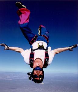
Pre-accident videos
Here are a few videos of Liam skydiving & landing before the accident. After viewing Liam’s landing style, it might be more apparent as to
why Liam ended up having the sort of accident that he did (it was enviable). At least he had a chance to enjoy skydiving before the accident.
[New images are surrounded by a RED border]
(Click on the small images to see larger versions or play a video.)
(For videos it is best to right click on the video, select "Save Target As..." and then save the video file to your hard drive to be played.)
[WARNING: Do not click on images marked with a red dot if you have a weak stomach]